


PreDocs
 JALLON Antoine - +33 489150861SEAL Tanush - +33 R
Engineers & Technicians
ROUQUET Sami - +33 489150866DELORME Barthélemy - +33 489150861HARRATHI Wafa - +33 489150861
Students
HOUESSOU Beni - +33 489150861
Masters
CHAOUQI Imane - +33 RKHROUFA Rania - +33 R
Visiting Professor
KARTTUNEN Mikko - +33 489150861
Do not miss this exciting talk from Professor Steffen LEMKE
When genes meet forces: revealing how mechanics helps shape the evolution of animal form. Do not miss this exciting talk ...
Read More
Read More
Join the latest MECABIONIC seminar presented by Professor Madzvamuse on new pattern formation theory
MECABIONIC Seminar Series Anotida Madzvamuse From: University of British Columbia 29th of April 2025 11:00, Meeting Room 5th floor Fizeau ...
Read More
Read More
Check out François Schweisguth exciting seminar on self-organized patterning in development
François Schweisguth From: Pasteur, Paris April 22 at 11:00, CB Conf. Dynamics of self-organized patterning in Drosophila Pattern formation in ...
Read More
Read More

Error correction in the developing embryo with Prof. Verena Ruprecht
Prof. Verena Ruprecht will give an exiting course open to all without registration at 8:30AM on Wednesday 26th of March ...
Read More
Read More
Mecanobiology course with Prof. Emmanuel Farge and Prof. Daniel Riveline
As part of the MECABIONIC program, Prof. Emmanuel Farge (Institute Curie) and Prof. Daniel Riveline (IGBMC, Strasbourg) will give a ...
Read More
Read More
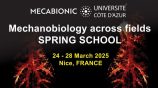
MECABIONIC Spring School: a theoretical and practical interdisciplinary workshop at UniCA
As part of the thematic semester MECABIONIC on the mechanobiology at Université Côte d’Azur, this spring school will bring together ...
Read More
Read More
Nuclear position takes central stage in tissue morphogenesis
Tissue morphogenesis is the change in shape of epithelia during embryo development and is key to give form and function ...
Read More
Read More

Mikko Karttunen (Western University, London, Canada) joins RAUZI team thanks to the IdEx Advanced Research Program 2024
Mikko Karttunen (Professor at the Western University, London, Canada) joins the iBV to work on a two-year collaboration project in ...
Read More
Read More

Do not miss an exiting talk by Prof. Edwin Munro from University of Chicago on epithelial zippering during neural tube formation
Dynamic origins of a self-pulled epithelial zipper We are using ascidian embryos as a simple model to understand the dynamics ...
Read More
Read More
Do not miss an exiting talk by Sylvain Gabriele from University of Mons on epithelia sensing curvature and spatial confinement
MECABIONIC Seminar Series More details ...
Read More
Read More