A morphogenetic wave travels along a biochemical guide to sculpt an epithelial furrow during embryo development
Epithelial furrowing is a fundamental morphogenetic process during gastrulation, neurulation and the shaping of the animal body. Epithelial furrowing often takes place in a stereotypic fashion: a fold forms at one position of a strip of tissue and eventually propagates along the tissue plane, following a linear trajectory. How fold formation and propagation are controlled and driven is still poorly understood. The Rauzi team, in collaboration with scientists at the faculty of mathematics and physics at the University of Ljubljana, has unveiled a morphogenetic wave-like process responsible for tissue furrowing. While the furrow location and initiation point is genetically predetermined, furrow propagation is driven by a mechanical wave sculpting the furrow. The wave relies on cell-cell interaction and it is generated by a group of cells functioning as a pacemaker triggering a domino effect.
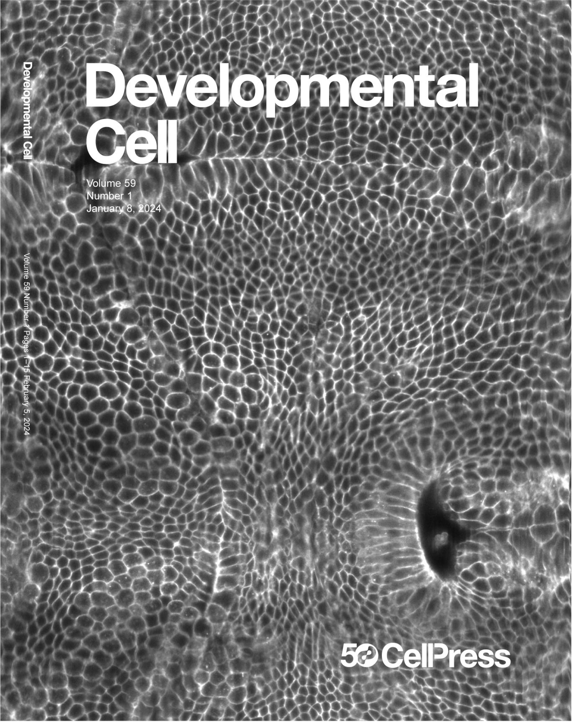
A furrow is a fold that develops along a line. Epithelial furrowing is a fundamental process during embryo development and organ formation. Furrow formation failure can result in severe neonatal disorders or even in a fully aborted development. Therefore, it is key to understand the fundamental mechanisms controlling and driving furrow formation to eventually apprehend the causes of furrow formation failure. To study this process, Popkova et al. have used the fruit fly embryo as a model system that provides an extremely powerful platform to apply live imaging and advanced genetics and molecular techniques. More specifically, the authors have focused their attention on the formation of the cephalic furrow (CF): a fold that travels along the dorsal-ventral axis of the animal and that separates the head from the trunk region of the embryo.
Popkova et al. show that intrinsic mechanical forces, generated by the actomyosin cytoskeleton within the CF region, are responsible for tissue folding. The furrow is then locked in place by de novo adherens junctions between cells facing each other within the fold. The authors show that the CF results from the propagation of cell shape transformation along a linear trajectory resulting in a morphogenetic wave. The position of the CF and the furrow initiation point are under the crossed control of the embryo anterior-posterior and dorsal-ventral gene patterning systems. Therefore, a morphogenetic wave propagates along a bi-dimensional genetic guide sculpting the furrow. Furrow formation is initiated by a group of cells functioning as a pacemaker and triggering morphogenetic changes that are cell-cell interaction dependent. This results in a domino effect (i.e., a trigger wave) that can be interrupted if a defect or mechanical barrier is introduced along the propagation path.
Finally, this study provides key information on how epithelial furrows are sculpted during embryonic development, opening new avenues to better decipher the causes of severe developmental disorders resulting from furrow formation failure. This knowledge has the potential to be used in the near future to engineer effective strategies to synthetically build and shape functional organs.

FIGURE LEGEND. Mercator projection of a living Drosophila embryo during gastrulation when morphogenetic waves sculpt folds and furrows in the blastoderm epithelium. Left is anterior, right is posterior, top is dorsal and bottom is ventral.
Link to the publication: https://t.co/vaJu2OffvV

