Imaging Facility

PRISM

Imaging Facility

Presentation

The imaging facility PRISM (Platform of Resources in Imaging and Scientific Microscopy) has been created to develop and provide access to state-of-the-art microscopes and together with expertise in optical microscopy and image analysis.
Fully integrated in the iBV on Valrose campus, the platform coordinates the imaging needs and provides expertise for research groups using a wide variety of biological models including cultured cells, yeast (S. cerevisiae and C. albicans), worms (C. elegans), flies (D. melanogaster), zebrafish, and mice, to address a range of fundamental scientific questions using different approaches.


The Imaging facility is also accessible to external users. Within MICA - Microscopy and Imaging Côte d'Azur - , PRISM is part of a joint imaging facility for 6 institutes which has been labelled by the GIS IBiSA. Based on this partnership, these different facilities coordinate their development to provide the largest panel of advanced microscopy techniques. Hence, PRISM facility is open to a wide variety of academic and industrial partners in the scientific community.

PRISM facility is part of the national networks RTmfm and the GDR ImaBio. Thus the platform is in contact with other facilities to exchange knowledge and tools about state of the art microscopy technologies.

Wide field

  • Axiocolor : AxioPlan Zeiss / Color camera for coloration
  • Axiofluo : AxioPlan Zeiss / Black and White camera for fluorescence


Video microscope

  • Videobserveur : AxioObserver - Zeiss (2011) / EMCCD ANDOR iXON 897 camera and sCMOS ANDOR Neo camera / MetaMorph software
  • Timelaps: Axiovert 200M - Zeiss (2004) / sCMOS Teledyne Photometrics PRIME BSI / MetaMorph software


Laser Scanning Confocal

  • Leica TCS SPE Microscope (2007)
  • Leica SP5 Microscope (2011): Hybrid detector / Galvanometric stage / resonant scanner
  • Zeiss LSM710 (2012): spectral detector / sensitive external detector (GaAsp)
  • Zeiss LSM780 (2011) : sensitive spectral detector (GaAsp) / Biphoton laser (MaiTai) / NDD detector in fluorescence and transmission
  • Zeiss LSM880 (2018) : sensitive internal detector (GaAsp) / Fast AiryScan detector / Piezo stage


Spinning Disk

  • Spinning disk: Olympus/Andor/Yokogawa X1 (2010) : Dual cam module (EM-CCD) / Fast 3D (piezo stage)
  • SPIN-FRAP-TIRF: Nikon TiE/Yokogawa W1 (2016) : photomanipulation module (Ilas2) / Andor iXon Life 888 (emCCD) / Fast 3D (piezo stage)


Super-resolution

  • Andor SRRF module on SPIN-FRAP-TIRF (2023)
  • Zeiss Elyra 7 (2023) : fast and resolutive Lattice-SIM microscopy


Lightsheet

  • Zeiss Lightsheet 7 (2023): dual side illumination for large and fast 3D imaging


Image analysis

  • Imaris for 3D analysis
  • Hygens for deconvolution


Engineers

Baptiste Monterroso
Tel : +33 489150781
E-mail : Baptiste.MONTERROSO@univ-cotedazur.fr

Sameh Ben Aicha
Tel : +33 489150780
E-mail : Sameh.BEN-AICHA@univ-cotedazur.fr

Scientific Coordinators

Robert Arkowitz
Tel : +33 489150740
E-mail : Robert.ARKOWITZ@univ-cotedazur.fr

Maximilian Fürthauer
Tel : +33 489150835
E-mail : Maximilian.FURTHAUER@univ-cotedazur.fr

iBV - Institut de Biologie Valrose

"Centre de Biochimie"

Université Nice Sophia Antipolis
Faculté des Sciences
Parc Valrose
06108 Nice cedex 2

iBV - Institut de Biologie Valrose

"Sciences Naturelles"

Université Nice Sophia Antipolis
Faculté des Sciences
Parc Valrose
06108 Nice cedex 2

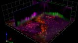
“Multi-angle-TIRF” : a molecular resolution optical microscope developed at iBV

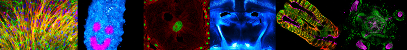
Being able to resolve biological signals and structures is central to understanding how molecules, organelles and cells are organized and ...
Read More