Adipo-Cible, a fundamental - translational - clinical research consortium on obesity co-sponsored by iBV! 30/08/2024 by Jean-Claude SCIMECA
Tenure-track position on “Quantitative Approaches for the Study of Regeneration” available at the Institut de Biologie Valrose, Nice, France 05/07/2024 by Jean-Claude SCIMECA
Guillaume Sandoz receives prestigious grant from the Simone and Cino Del Duca Foundation 27/06/2024 by Jean-Claude SCIMECA
REBICA 2024 - REncontre annuelle BIoinformatique à l'Université Côte d'Azur 26/06/2024 by Jean-Claude SCIMECA
Do not miss an exiting talk by Sylvain Gabriele from University of Mons on epithelia sensing curvature and spatial confinement 30/04/2024 by Jean-Claude SCIMECA
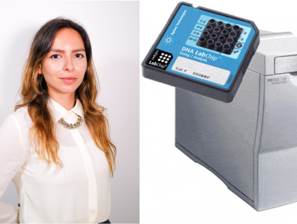
Spotlight on the study published by Michèle Studer in the journal Protein Science in partnership with the "genetic code expansion" (GCE) platform headed by Krittalak Chakrabandhu 15/04/2024 by Jean-Claude SCIMECA