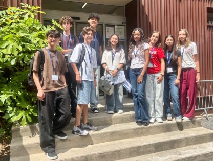
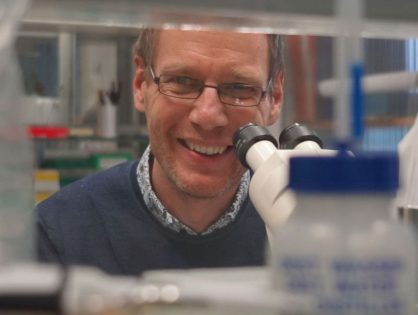
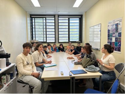
“Amazing, incredible, so much fun!”: Nine high school students immerse themselves in research at iBV 29/06/2026 by Jean-Claude SCIMECA
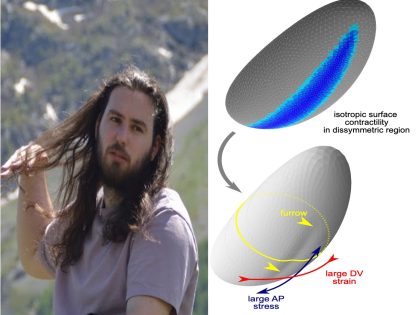
‘Active Viscoelasticity and Shell Mechanics in Morphogenesis’, with Antoine Jallon 23/06/2026 by Jean-Claude SCIMECA
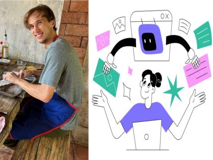
’GPTs in Biology: Genius Assistant or Confidently Wrong Lab Partner?’, with Kristóf Kovacs 17/06/2026 by Jean-Claude SCIMECA
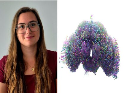
“Exploring Brain Asymmetry Through the Drosophila Connectome”, with Coralie Mouchet 17/06/2026 by Jean-Claude SCIMECA
Poste d’Ingénieur d’Études CNRS en Expérimentation et Instrumentation Biologiques 10/06/2026 by Jean-Claude SCIMECA
“From Gene to Organ” outreach program awarded the Scientific Rigor Prize! 21/05/2026 by Jean-Claude SCIMECA
Protocol for preparation of a synaptosome-enriched fraction to profile presynaptic RNA content in adult Drosophila brains 06/05/2026 by Jean-Claude SCIMECA
Genetic code expansion: Bridging synthetic biology precision and cellular complexity to decipher molecular mechanisms of diseases 04/05/2026 by Jean-Claude SCIMECA