The egg puts on a corset to get the right shape

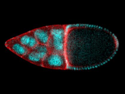
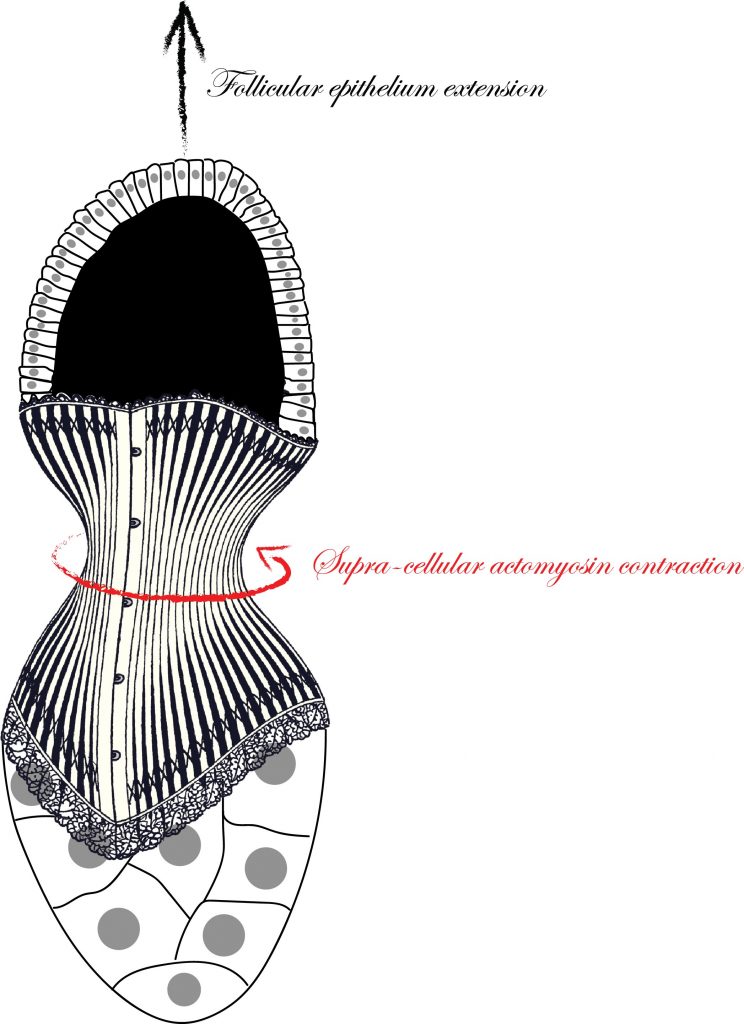
Thanks to an interdisciplinary and exciting collaboration between the iBV and the CBI in Toulouse, the Rauzi and the Wang teams unveiled a Cdc42 dependent mechanical process responsible for tissue elongation in the developing Drosophila egg chamber. By engineering new optogenetic techniques and infra-red laser based surgery, Anna Popkova and others show that a polarized supracellular actomyosin network, working as a molecular corset, generates tissue scale forces directing egg chamber elongation. Finally, this study opens new avenues to better understand how supra-cellular cytoskeletal networks emerge to drive embryo-scale morphogenesis during development. This work was published in Nature Communications.

Understanding the mechanisms that are responsible to shape tissues and organs is an important and exciting challenge in developmental biology. The actomyosin cortex often plays a key role in generating the forces necessary to shape individual cells. Nevertheless, the actomyosin architecture can extend beyond the size of a single cell: cytoskeletal networks emerge at the scale of a tissue. These supra-cellular contractile structures can generate large forces that can rapidly shape epithelia.

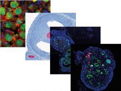
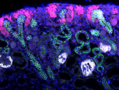
In this study, scientists from the Rauzi and the Wang lab use the Drosophila egg chamber as a model system to study the origin and the mechanics of supra-cellular actomyosin networks. Popkova and others show that a supra-cellular array of parallel actin bundles, emerging from interdigitating filopodia, envelopes the follicle tissue. Filopodia radiate in a polarized way from basal stress fibers and extend by penetrating the neighboring cell cortexes. Filopodia can be mechanosensitive and function as anchors between cells. The small GTPase Cdc42 governs the formation of intercellular filopodia and stress fibers in the follicular cells. Thus, a Cdc42-dependent supracellular cytoskeletal network provides a scaffold integrating local oscillatory actomyosin contractions at the tissue scale to drive global polarized forces and tissue elongation.

Inset

Actin filaments assemble into diverse protrusive and contractile networks to generate forces in diverse cellular processes. Stress fibers are contractile higher order cytoskeletal structures composed of actomyosin bundles. Stress fibers play a key role in generating forces along the fiber direction and have important implication in cell adhesion to the extracellular matrix.

Filopodia are dynamic, finger-like plasma membrane protrusions that act as antennae to sense the mechanical and chemical environment. They are often regarded as “sensory organelles”. Filopodia are involved in many biological processes, such as growth cone guidance, cell migration, wound closure, and macrophage-induced cell invasion. These thin membrane protrusions are 60–200 nm in diameter and contain parallel bundles of 10–30 actin filaments held together by actin-binding proteins. 

Laser dissection is a useful tool in developmental biology to probe the mechanical forces from the subcellular to the tissue/embryo scale. During tissue morphogenesis, cells are equipped with actomyosin networks generating forces. In this study, researchers used near-infrared (NIR) femtosecond (fs) pulsed laser surgery to dissect the actomyosin cytoskeleton with subcellular precision.  This technique allows to selectively ablate actomyosin networks while preserving the cell plasma membrane. The resulting recoil of the remaining network, after laser dissection, is imaged and analyzed to deduce local forces responsible for tissue morphogenesis.

Optogenetics allows to control, via light stimulation, protein conformation changes and thus protein activity with spatial and temporal specificity. By using molecular engineering, proteins of interest are fused to photo-activatable proteins than can be expressed in specific cells. Using the optogenetic tool PA-Cdc42 which leads to the expression of a light-activatable Cdc42 protein, researchers were able to precisely determine the role of Cdc42 in follicular cells.

To read more: 

A Cdc42-mediated supracellular network drives polarized forces and Drosophila egg chamber extension.
Popkova A, Stone OJ, Chen L, Qin X, Liu C, Liu J, Belguise K, Montell DJ, Hahn KM, Rauzi M@, Wang X@.
Nat Commun. 2020 Apr 21;11(1):1921. doi: 10.1038/s41467-020-15593-2.


Press release : Actualités scientifiques de l’INSB

Movie: Time-lapse of a representative mCD8GFP-expressing egg chamber labelled with MyoII-mCherry. Laser dissection of the supra-cellular actomyosin network was performed along the AP axis of the egg chamber. Scale bar 10 μm.

PhD Position - 3 years - Starting October 2020

Deciphering the regulation of the cell death receptor Fas by cell polarity molecules & adherens junctions in both tumoral and normal human epithelia

Key words: Receptor, Signaling, Cancer, Cell polarity, Cell-Cell jonctions


The PhD student will integrate the research group “death receptors signaling in cancer therapy” (iBV, http://ibv.unice.fr/research-team/hueber). at the Institute of Biology Valrose affiliated to the CNRS, Inserm and University Côte d’Azur (UCA).

Project proposal: Our team is investigating the functions of the cell death receptor Fas/CD95/TNFRSF6, a member of the TNFR superfamily. Fas is considered as a tumor suppressor thanks to its ability to eliminate cancer cells by engaging programmed cell death by apoptosis. However, Fas activation by its ligand (FasL) could also promote tumor development and immune disorders (1). Our group is studying the molecular mechanisms that control the Fas versatile signaling outcome in the context of both normal and cancer cells (2, 3). Our recent data show that formation of adherens junction, a cell-cell adhesion complex, and association with the Dlg1 polarity complex prevent the pro-apoptotic signaling of Fas (4, 5). This new Fas-regulatory mechanism is crucial to protect normal epithelial cells from apoptotic signals and to sense and eliminate abnormal cells from epithelial tissues to prevent pathological outcome such as cancer and chronic inflammatory diseases. The PhD student will pursue this project and decipher the regulation of Fas cell death and non- death signaling by the Cadherin-Dlg1 polarity complex notably by studying Fas receptor signaling/trafficking on both primary and tumoral epithelial human cells by using various cell biology approaches.

Bibliography of the team link to the project: 1- Rossin et al (2019) Cancers, 8;11(5):639. 2- NL t al (2018) Sci Rep, 20;8(1):12424. 3- Chakrabandhu K (2016), PLoS Biol, 4;14(3). 4- Gagnoux-Palacios, L.; et al (2018) Journal of Cell

Biology, 217, 3839-3852. 5- Gagnoux-Palacios L., Hueber AO (2019) Medecine/Sciences. 35(11):830-833.

Technical approaches: Human cell culture; cell death assays; receptor trafficking studies (cell surface labeling, endocytosis assay), protein expression quantification/localization (FACS, IP, IF, IB, ELISA, proteomic); microscopy techniques (confocal imaging, time-lapse).

Candidate profil: We are looking for a highly motivated student, independent and creative, with a Master’s Degree in Cellular, Cancer or Molecular Biology. Prior experience with cell culture and classical cellular and biochemical approaches will be appreciated.

HOW TO APPLY: Interested and motivated students should send as soon as possible a CV, a motivation letter, master scores/ranking and reference letters to both L. GAGNOUX (gagnoux@unice.fr) and A-O HUEBER (hueber@unice.fr).

1 Doctoral position - Deadline June 1st, 2020 (Optogenetics)

 Doctoral position at University Côte d’Azur: Fungal Cell Biology

Light-dependent regulation of cell polarity in a fungal pathogen

The fungus Candida albicans is normally a harmless commensal that is found on mucosal surfaces of the gastrointestinal and urogenital tract in most healthy individuals. This commensal organism can cause superficial as well as life-threatening systemic infections in response to alterations of its environment, and is particularly aggressive in immuno-compromised individuals. As an opportunistic pathogen it can colonize and infect different body sites and is responsible for one of the most predominant fungal nosocomial infections. The ability of this fungus to switch from an ovoid form to a filamentous form is critical for its pathogenicity, in particularly its ability to invade and penetrate into host tissues and evade and burst out of host immune cells.

The recent advent of light-dependent approaches to control protein subcellular localization has made possible the specific alteration of growth, circumventing classical genetic and chemical perturbations. We have optimized such a light-dependent protein targeting system for C. albicans, which gives us exquisite control of growth in this organism. In addition, new variants of these systems have been established which facilitate their use and response time. Furthermore, a number of new, spectrally distinct fluorescent proteins, which we have optimized for use in C. albicans, now make it possible to follow different cellular processes simultaneously during light-dependent perturbation of growth and cell polarity. The goal of this project is to use such a light-dependent protein targeting to probe filamentous growth as well as invasive filamentous growth in a human fungal pathogen. The project will use a combination of molecular biology, microbiology and live cell microscopy to probe filamentous growth and morphogenesis in this fungal human pathogen.

We are seeking highly motivated candidates with a background in Cell Biology and interest in live cell imaging. Experience in Microbiology would be a plus.

Interested candidates can contact R. Arkowitz (arkowitz@unice.fr) by June 1st


1) M Bassilana, C Puerner & RA Arkowitz. Curr. Opin. Cell Biol. 2020 62:150-158.
2) PM Silva, C Puerner, A Seminara, M Bassilana & RA Arkowitz. Cell Rep. 2019 28:2231–2245.
3) RA Arkowitz & M Bassilana. F1000 Res. 2019 8.
4) A Weiner, F Orange, S Lacas-Gervais, K Rechav, V Ghugtyal, M Bassilana & RA Arkowitz. Cell Microbiol. 2019 21: e12963
5) H Labbaoui, S Bogliolo, V Ghugtyal, NV Solis, SG Filler, RA Arkowitz & M Bassilana. Plos Pathog. 2017 13: e1006205


1 Doctoral position - Deadline June 1st, 2020 (SuperRes)

Doctoral position at University Côte d’Azur: Fungal Cell Biology

Organelle dynamics in a human fungal pathogen at high temporal and spatial resolution

Worldwide, fungal infections cause significant morbidity and mortality and Candida species are the major etiological agent of such life-threatening infections and represent an emerging global microbial threat. A range of advances in medical treatment have increased life expectancy yet also dramatically increased the population of elderly as well as severely ill patients, highly susceptible to nosocomial infections, in particular those caused by fungi such as Candida albicans. C. albicans is normally a harmless commensal, found on mucosal surfaces of the gastrointestinal and urogenital tract in most healthy individuals that causes superficial as well as life-threatening systemic infections in response to alterations of its host environment. It is particularly aggressive in immuno-compromised individuals. The ability of this organism to switch from an ovoid to a filamentous form, concomitant with changes in cell surface antigens and enzyme production, is critical for its pathogenicity, in particular to invade host tissues and evade host immune cells. Many fungi, including C. albicans form elongated hyphal filaments that are tube-like cells in which growth is restricted to the tip. This dramatic yeast to filament cell shape change is a distinct advantage for studying the regulation of cell polarity and membrane traffic, critical for such morphogenesis. However, the hyphal apical zone is densely packed, with multiple membrane compartments including secretory vesicles below the light resolution limit and somewhat larger Golgi cisternae in a small volume at the filament tip and these compartments are highly dynamic making live cell imaging extremely challenging, in particular with high temporal and spatial resolution.

To understand the exquisite regulation of tip growth, the aim of this project is to quantitate the movement of membrane compartments in 3D with high spatial and temporal resolution. Conventional methods including fluorescence microscopy and electron microscopy suffer from either limited spatial or temporal resolution, whereas of super-resolution microscopy approaches, has made imaging with a resolution higher than that imposed by the diffraction limit of light possible. This interdisciplinary project (carried out with Laure BLANC‐FERAUD, I3S Laboratory – UMR CNRS 7271) will take advantage of fluorescent molecule blinking and their fluctuations over time, to generate super-resolved images with high temporal resolution. The goal of this project is to develop, optimize and apply such methods, specifically adapted to a multimodal microscope established at the iBV, to the investigate the reorganization of the membrane compartments during C. albicans filamentous growth, with high temporal and spatial resolution. This project will involve extensive state-of-the-art microscopy as well as optimization of image reconstruction and analyses computational algorithms.

We are seeking highly motivated candidates with a background in Cell Biology and interest in live cell imaging. Experience in Microbiology would be a plus.

Interested candidates can contact R. Arkowitz (arkowitz@unice.fr) by June 1st


1) M Bassilana, C Puerner & RA Arkowitz. Curr. Opin. Cell Biol. 2020 62:150-158.
2) PM Silva, C Puerner, A Seminara, M Bassilana & RA Arkowitz. Cell Rep. 2019 28:2231–2245.
3) RA Arkowitz & M Bassilana. F1000 Res. 2019 8.
4) A Weiner, F Orange, S Lacas-Gervais, K Rechav, V Ghugtyal, M Bassilana & RA Arkowitz. Cell Microbiol. 2019 21: e12963
5) H Labbaoui, S Bogliolo, V Ghugtyal, NV Solis, SG Filler, RA Arkowitz & M Bassilana. Plos Pathog. 2017 13: e1006205