PhD student - Nuclear migration and packing for the emergence of a multicellular system - Beginning on the 1st of October 2026 09/04/2025 by Jean-Claude SCIMECA
Error correction in the developing embryo with Prof. Verena Ruprecht 25/03/2025 by Jean-Claude SCIMECA
Mecanobiology course with Prof. Emmanuel Farge and Prof. Daniel Riveline 21/03/2025 by Jean-Claude SCIMECA
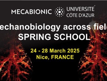
MECABIONIC Spring School: a theoretical and practical interdisciplinary workshop at UniCA 20/03/2025 by Jean-Claude SCIMECA
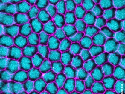
Nuclear position controls the activity of cortical actomyosin networks powering simultaneous morphogenetic events 13/02/2025 by Jean-Claude SCIMECA